Library search
Search Results
-
 Possibilities of mini-invasive diagnostic and treatment methods in closed abdominal injuries
Catalog of dissertations and abstractsThe aim of the study is to improve the results of diagnosis and surgical treatment of victims with closed abdominal injury by developing a new approach to ultrasound assessment of the amount of hemoperitoneum, expanding and specifying indications for laparoscopy, taking into account the volume of free fluid in the abdominal cavity.
The object of the study were 160 patients with closed abdominal injury with stable hemodynamics, was hospitalized in the surgical Department of the Republican specialized scientific and practical center for emergency medicine of the Samarkand branch (clinical departments of surgical diseases № 2 and surgery postgraduate faculty of Samarkand state Medical Institute) for the period from 2010 to 2019.
The scientific novelty of the study is as follows: a fundamentally new approach to ultrasound evaluation of discrete volumes of free fluid in the abdominal cavity is proposed, based on taking into account the thickness of the fluid layer and its prevalence in the abdominal cavity zones; The expediency of using the ultrasound indicator "free fluid in the abdominal cavity < or >500 ml" in choosing the tactics of surgical treatment of patients with closed abdominal injury is substantiated; an algorithm for choosing surgical tactics for the treatment of patients with closed abdominal trauma was developed based on an ultrasound assessment of the volume of free fluid in the abdominal cavity.
Implementation of research results. Based on the results of a scientific study to improve the diagnosis and surgical treatment of patients with closed abdominal trauma:methodological recommendations "The choice of tactics for surgical treatment of closed abdominal trauma based on ultrasound assessment of the nature and severity of the injury" have been developed (certificate of the Ministry of Health No. 8n-z/1282 dated November 15, 2022). The proposed recommendations made it possible to increase the effectiveness of the diagnosis of intra-abdominal injuries in patients with abdominal trauma;
The results of scientific research on improving the diagnosis and surgical treatment of patients with closed abdominal injury have been introduced into medical practice, including the clinical practice of the Republican Scientific Center for Emergency Medical Care and its Samarkand, Surkhandarya and Navoi branches (conclusion of the Ministry of Health No. 8 n-z/699 dated December 21, 2022). The introduction of the obtained results into clinical practice allowed to improve the quality of high-tech surgical care provided to patients with isolated and combined abdominal injuries, to reduce the frequency of postoperative complications from 11.9 to 3.1% (p=0.144).
The structure and volume of the dissertation. The dissertation consists of an introduction, 4 chapters, conclusions and a list of cited literature. The volume of the text material is 107 pages. -
 CLINICAL AND ANAMNESTIC CHARACTERISTICS OF PATIENTS WITH CHANGES IN THE ORGAN OF VISION IN ATHEROSCLEROSISTo study the clinical and anamnestic characteristics of patients with changes in the organ of vision in atherosclerosis (AS). Material and methods: 156 patients with laboratory-clmically diagnosed atherosclerosis underwent examination and dynamic observation. All patients underwent a comprehensive examination, including the study of central visual acuity, kinetic and computed static perimetry, tonometry, gonioscopy. biomicroscopy, fundus ophthalmoscopy, ultrasound Doppler ultrasound of the vessels of the eye and the brachiocephalic trunk. Results: In patients with initial atherosclerosis in the absence of any subjective complaints and clinical manifestations of atherosclerotic vascular lesions, with a careful survey, episodes of short-term visual impairment after overeating, physical overstrain, visual impairment in one eye in bright light, periodic fog m front of the eye. discomfort, etc. dry eye. Patients with a clinical course of atherosclerosis, patients noted decreased vision, disappearance of the lower half of vision, upper half of vision, floating spots m front of the eye. pain in the eye. fog in front of the eye and loss of visual acuity, etc. Conclusions: Taking into account the presence of both modifiable in patients, and unmodifiable risk factors for the development of atherosclerosis, early and differential diagnosis of atherosclerosis should be carried out to prevent damage to target organs.
Stomatologiya -
 MOTIVATIONAL ASPECT OFIMPROVING MOUTH HIGIENE OF THE CHILDREN DURING ORTHODONTIC TREATMENTЛ comparative assessment of the health status of organs and tissues of the oral cavity was conducted, based on the dynamic monitoring of the dental status of 42 children (15 boys and 27 girls) aged 6 to 12 years, who had undergone the orthodontic treatment using myofunctional devices. Children supervision was carried out during 3 months. The patients were divided into 4 groups after finishing an oral cavity sanitation. Children of three groups, who had been given one of three types of R.O.C.S, toothpastes, which they had chosen themselves, improved their oral hygiene indexes for three months. Children who hadn't been given the toothpaste, but they had got the advices of hygiene of an oral cavity, improved their oral health indexes less than children from the other groups. The patients came for the re-inspection every month. It is proved that usingof toothpastes R.O.C.S. Junior visiblyimproves the hygiene and oral health of children aged from 6 to 12 years, whichever paste they choose.
Medicine and innovations -
 The role of QLF technology in improving oral hygiene and behavior to preserve oral health in childrenThe aim of the study was to determine oral hygiene, oral health behaviors of children and motivate to improve them by using portable QLF device such as Qscan. One hundred children aged 14-16 years were included in the study. The children were divided into 2 groups. To the control group of children hygienic lessons were provided as a traditional lecture-training (10 min.). The experimental group were provided by a traditional method combined with the demonstration of plaque level using Qscan device. After 2 weeks of examination both groups showed better significant changes in oral hygiene and oral health knowledge, behavior and attitude. However at the end of trial after 2 months oral hygiene and oral health knowledge indices showed significant better changes in experimental group (0.07±0.02: 29, respectively), however no significant changes were discovered m a control group (0.46±0.04: 19). It can be concluded that Qscan aided oral hygiene education was usefill to train children and showed better and long lasting results.
Stomatologiya -
 FEATURES OF THE MICROECOLOGY OF THE ORAL CAVITY IN PARTIAL SECONDARY ADENTIAAccording to the World Health Organization, one of the common dental diseases is partial secondary adenatia. By distribution, this pathology affects 75% planet population. The defects of the dentition affect not only the state of the organs of the maxillofacial region, but also on the condition of the whole organism, and therefore leads to a violation of homeostatic equilibrium. The combination of microorganisms in the oral cavity is a stable biocenosis, with a certain species composition, which can be modified qualitatively or quantified in accordance with the type of oral disease. The microflora of the oral cavity is distinguished by a variety of species persistently react to endo and exogenous factors. With partial loss of teeth, the microflora of the oral cavity is changing, as well as one of the destabilizers of the biocenosis of the oral cavity arc prosthetic structures.
Medicine and innovations -
 VIOLATIONS OF MICROECOLOGY AND LOCAL FACTORS OF PROTECTION OF THE ORAL CAVITY IN PATIENTS WITH VIRAL HEPATITIS BThe normal microflora of the oral cavity is a biological barrier that prevents the reproduction of random and pathogenic flora. In addition, it serves as a constant stimulator of local immunity. It is known that the development of pathology in the oral mucosa can be influenced by diseases of the internal organs, including the liver and hepatobiliary system. An important discovery was the establishment of the possibility of extrahepatic replication of viruses of parenteral hepatitis "B" and "C", in particular, in the cells of the bone marrow, blood, lymph nodes and spleen. Viral lesions of the liver with extrahepatic replication of viruses cause immunodeficiency states in the body, which, as a rule, are also reflected in local oral cavity protection factors. Aims: to study the quantitative and qualitative composition of the microflora of the oral cavity and the indicators of local protective factors in patients with viral hepatitis B. Material and methods: microbiological and immunological studies were carried out in 142 patients with chronic viral hepatitis B. Results: The development of a generally immunodeficient state in the oral cavity of patients with chronic viral hepatitis B contributes to a decrease in both cellular and humoral immunity, and leads to a microbial overgrowth syndrome, that is, a dysbiotic state in the oral cavity in these patients.
Medicine and innovations -
 The prevalence of mycotic infections reaches 40 — 88%. Questions of early effective diagnostics, pathogenesis, therapy and prevention of this pathology in many respects remain unresolved, debatable though numerous both clinical, and experimental works are devoted to their studying. By authors it is studied the frequency and nosological forms of candidiasis connected with age of patients. At the same time the main specific weight in a total amount of diseasesrepresents at the age period from 34 to 64 yearsand older sick people. The high incidence of adult population is explained by higher frequency of background somatic pathology, frequent reception of antibiotics and hormonal (steroid medicines).
 -
 The role of magnetic resonance imaging in the comprehensive radial diagnosis of volumetric masses of the eye organ
Catalog of abstractsRelevance of the problem. The difficulties of diagnostics of orbital diseases are well known. Especially difficult is intraspecies differentiation among the multitude of tumour, pseudotumour, inflammatory, vascular, endocrine and other diseases occurring here, manifested by the symptom complex of unilateral exophthalmos [Beradze I.N., 1978; Brovkina A.F., 1993].
Malignant intraocular neoplasms are the main cause of death of patients with diseases of the organ of vision, with 45-48% of patients dying from metastases in the first 5 years after enucleation [Alekseeva I.B., 1990, Barkhash S.A.1978, Brovkina A.F..1991, 1997; Keizer R.W.. Viclvoyc G.L.,1986],
Retinoblastoma is the most frequent malignant neoplasm in children. According to different authors, the frequency of its occurrence is 1 case per 14000 - 35000 newborns. [Bobrova N.F. and Vit V.V., 1993; Brovkina A.F., 1997; Provenzale J.M., et al., 1995; Skulski M., et al., 1997; Weber A.L., Mafee M.F, 1992; Wilms G., et al., 1989]. The frequency of patients with the most malignant intraocular tumour in adults - uveal melanoma has recently reached 7-9 people per 1 million population [Brovkina A.F., 1997; Kotslyansky E.O., 1989; Yushko N.A., Peskova L.I., Kalenich L.A., 1989; Peyster R.G., Augsburger J..I., Shields J.A., 1988; Romani A.. Baldeschi L., ct al 1998; Scott I.U., 1998].
The fundamental difference in treatment tactics, depending on the stage of development, size and topography of the tumour, as well as the seriousness of the prognosis in retinoblastomas and melanomas sharply increase the requirements for the accuracy of their differential diagnosis. At the same time, the number of diagnostic errors in ocular tumours continues to be 10-30% even when complex clinical and instrumental examination is applied in specialised ophthalmological centres [Ternovoy S.K., Panfilova G.V., Rogozhin V.A., 1979; Friedman F.E., Malyuta G.D., Kodzov M.V., 1995; Song G.X., 1991].
Widely used in ophthalmological practice traditional diagnostic methods (ophthalmoscopy, gonioscopy, diaphanoscopy, fluorescence angiography, laboratory tests) are insufficient to obtain comprehensive information about the localisation, nature of growth and prevalence of volumetric pathological formations of the eye and orbit. This circumstance and not quite satisfactory results of surgical treatment are the causes of high mortality of patients [Muratova T.T., Nigmanova N.H., Kozlovskaya G.M.. 1989, Naches A.I., 1980; Cheremisin V.M., Trufanov G.E., Kholin A.V., 1991]. Untimely or erroneous recognition of pathological processes of the orbit leads to a sharp deterioration of visual functions, up to blindness, and in some cases to the death of the patient [Yuzhakov A.M., Travkin A.G., Kiseleva O.A., 1991]. All this determines the importance of timely and accurate diagnosis of diseases of the orbit, on the one hand, and the difficulty of such diagnosis - on the other [Gabunia R.I., Kolesnikova E.K., Tumanov L.B., 1982].
The fact that the orbit is closed from direct inspection and palpation by bone walls and the eyeball, indicates the advantage of radial diagnostics in comparison with other methods of examination. In the arsenal of clinicians there is a great variety of methods of clinical-radial diagnostics of orbital pathology, however, at present the information in the literature about their resolving capabilities and significance in comparative aspect is incomplete and not fully studied. The priority of using one or another instrumental investigation, their sequence and expedient combination have not been determined yet. This makes it difficult to choose the optimal standardised approach for diagnosis and adequate treatment [Cheremisin V.M., Trufanov G.E., 1993, Weber A.L., Sabates N.R., 1996; Wenig V.M., Mafee M.F., 1998].
Thus, the study of these and other questions, contributing to the improvement of diagnostics and treatment of patients with neoplasms of the eye and ocular cavity, should be recognised as urgent urgent.
Purpose of the study. Comparative evaluation of magnetic resonance tomography capabilities and development of algorithms for complex radial diagnostics of volumetric formations of the visual organ. To solve this goal we set the following tasks.
1. To study the normal picture of the magnetic resonance image of the visual organ in comparison with other methods of visualisation.
2. To find out the possibilities of magnetic resonance tomography, ultrasound and computed tomography in detection and evaluation of intraocular neoplasms.
3. To determine the role and place of magnetic resonance tomography in differential diagnostics of volumetric pathological formations of the eye cavity in comparison with other radial methods of research.
4. To determine the indications and to develop an algorithm for the complex application of radiography, ultrasound, computer and magnetic resonance tomography for diagnostics of volumetric formations of the eye organ.
Scientific novelty.
The present work is the first to give a detailed and detailed description of the complex clinical and radiation examination, with generalisation and standardisation of magnetic resonance, computer and ultrasound semiotics of volumetric pathological formations of the eye and eye cavity. The conducted clinical and instrumental investigations allowed to determine the diagnostic value and resolving capabilities of each of the applied methods. The ultrasound, CT and MRI signs of volumetric formations of the eye organ were studied, clarified and supplemented taking into account the use of low-field magnetic field and general-purpose ultrasound apparatus. The developed standardised diagnostic algorithm of examination of patients with this pathology is new, thanks to which the pre-oppositional diagnosis of tumour and other diseases of the visual organ is improved and the total radiation load on the patient is reduced.
Conclusions
1. MPT will provide an opportunity to study the weight of the soft tissue and anatomical components of the ocular cavity, up to the optic nerve sheath and perineural liquor space, the orbital apex and chiasmal-sellar region, as well as to assess the condition of adjacent structures of the brain and facial skull. The method is limited in the evaluation of changes in the bony walls of the orbital cavity.
2. MRI is inferior in detecting characteristic signs of retinoblastoma (presence of calcification). The sensitivity of MRI was 66.6%, while for ultrasound and CT these values were 96.1 and 100%, respectively. But when the tumour spreads rstrobulbarly outside the eyeball (at 3-4 stages) the informativeness of MRI increases significantly. In uveal melanoma the sensitivity and specificity of MRI reaches 100%.
3. Both MRI and CT have a high detection rate (98.1% and 95.8% respectively) of benign orbital tumours of both primary and secondary origin. However, MRI is the preferred method of investigation. MRI is especially informative when a cranioorbital tumour and pseudotumour are suspected. The sensitivity of the method is 90.9% and 91.6%, respectively
4. In some cases ultrasound can be used to differentiate between encapsulated and diffuse neoplasms, which facilitates the diagnosis. However, when the pathological process is localised near the orbital apex, the diagnostic value of ultrasound decreases. In such cases it is advisable to use MRI.
5. In detection of primary and secondary malignant tumours of the orbital cavity both MRI and CT are quite informative (sensitivity 97,2% and 95,4% respectively), but the most comprehensive information about the state of bone walls will be provided by CT. When the process spreads intracranially, the value of MRI increases significantly, especially with the use of contrast enhancement.
6. The developed algorithm of complex clinical and radiation examination of patients with the use of ultrasound, CT and MRI is the most effective in the diagnosis of volumetric pathological formations of the eye and eye cavity, allowing to reduce to an adequate minimum the total radiation load on the patient and diagnostic period, excluding duplication of research techniques and choosing the most informative in each case, which in turn allows to develop appropriate treatment tactics and reduce the level of disability of the patient. -
 Особенности гигиены полости рта при лечении заболеваний пародонта и кариеса зубов
Actual problems of dentistry and maxillofacial surgery 4Достижение высокого эстетического результата невозможно без учета состояния гигиены полости рта и наличия заболеваний пародонта. От уровня гигиены полости рта и состояния тканей пародонта напрямую зависят качество и длительность функционирования реставраций. Достижение высокого эстетического результата невозможно без учета состояния гигиены полости рта и наличия заболеваний пародонта. От уровня гигиены полости рта и состояния тканей пародонта напрямую зависят качество и длительность функционирования реставраций. Однако индивидуальная гигиена полости рта, профилактика, ранняя диагностика и лечение заболеваний пародонта при лечении кариеса часто упускаются из внимания практикующих врачей
-
 Study of bacterial oral indexes in patients men seeking professional cleaning of oral cavity in SamarkandThe microflora of the oral cavity is considered as the primary target for any factor that directly or indirectly affects bat the adhesion of opportunistic symbionts and colonization of microorganisms. Objective: To study the bacterial flora of the oral cavity of Samarkand residents. Bacteriological research objects were not stimulated mixed saliva, plaque, swabs from the surface of fillings. Number of cariogenic microorganisms S. mutans, S. rattus in all samples from the oral cavity before readjustment exceeds 106, 107 CFU / ml. Qualitative analysis of oral microorganisms was completed by release of 260 strains of microorganisms belonging to 11 genera.
Journal problems of biology and medicine -
 MAST CELLS AND GLYCOSAMINOGLYCANS IN THE MUCOUS MEMBRANE ORAL CAVITY IN BEHCET'S DISEASEMast cells (MC) and glycosaminoglycans (GAG) in various zones of oral mucosa are investigated during remission and aggravation of Behcet disease (BD) in 10 patients (8 men and 2 women; average frequency of aggravations in a year - 4.5). Bioptats (biopsy material) of various zones of oral mucosa arc investigated histochcmically. The MC arc identified by 0.05 percent thioninc solution according to Hassanov's prescription, as well as by GAG-PAS-reaction under the control of amylase, and by fast blue and strong garnet CBS [ 1 J. The statistical correlation between frequency of aggravations of BD and histochemical quantitative indices of the MC and GAG is studied.
Medicine and innovations -
 New technologies for the prevention of adhesions in thoraco-abdominal surgery
Catalog of dissertations and abstractsThe aim of the research work is determination of the prospects for the use of a domestic agent for the prevention of adhesion formation in thoraco-abdominal surgery on the basis of experimental and morphological studies.
Research objectives were white outbred rats in the amount of 62 individuals, in two experimental studies on the abdominal and pleural cavities, in each series of experiments the studies were carried out in 2 comparative groups, control and main. Experiments on the formation of adhesions in the abdominal and pleural cavities were carried out on the basis of the Republican Specialized Scientific and Practical Medical Center of Surgery named after acad. V.Vakhidov in the Department of Experimental Surgery for the period from 2019 to 2020.
The scientific novelty of the research consists of the followings: it is proved according to the data of experimental research that when modeling the adhesion process in the abdominal cavity, the local application of an anti-adhesion coating made of cellulose derivatives reduces the processes of adhesiogenesis and the development of changes in architectonics, bends and narrowings of the intestinal lumen; it was found in an experimental study that when modeling the adhesion process in the chest cavity, the local use of an anti-adhesion implant provides a significant decrease in the risk of adhesiogenesis in the form of the formation of coarse adhesions or planar adhesions; it was determined that when blood serum was applied over a powder implant, the quality of adhesion and the uniformity of its distribution on the surface of the experimental defect of the peritoneum or lung did not change, but, in contrast to activation by blood (to ensure a hemostatic effect), it was not accompanied by the development of cellular inflammation due to the resorption of thrombotic masses; it was found that the formation of a gel film over the area of damage to the peritoneum in the absence of cellular elements of blood makes it possible to achieve biodegradation of the coating without a pronounced cellular-inflammatory reaction, providing cicatricial replacement of defects with a significant reduction in the risk of developing a massive adhesive process; the morphostructural features of the formation of the adhesive process when using an anti-adhesive coating, characterized by regression in the dynamics of the number of connective tissue cells of the inflammatory infiltrate with scarring of the defect zone without the development of adhesive conglomerates with the surrounding tissues, have been determined.
Introduction of the research results. According to the results of a scientific study on a comparative analysis of the use of a domestic agent for the prevention of adhesion formation in thoraco-abdominal surgery: methodological recommendations were developed: "New technologies for the prevention of adhesions in thoraco-abdominal surgery" (certificate of the Ministry of Health No. 08-09/10055 of August 12, 2021). The proposed recommendations for performing surgical interventions on the organs of the abdominal and thoracic cavities will allow for sparing local hemostasis, as well as prevent the formation of a coarse adhesive process in the abdominal cavity.
The obtained scientific results on a comparative analysis of the use of the domestic remedy for the prevention of adhesion formation in thoraco-abdominal surgery have been introduced into the practical activities of health care, including in the Republican Specialized Scientific and Practical Medical Center for Surgery named after V.I. Academician V. Vakhidov, surgical departments of the clinics of the Andijan and Samarkand State Medical Institutes (certificate of the Ministry of Health No. 08-09/10055 of August 12, 2021). Based on the proposed results of experimental studies, it was shown that the use of an anti-adhesive coating made of cellulose derivatives made it possible to reduce the risk of adhesion formation from 60% to 20%, bowel deformation without manifestations of obstruction from 33.3% to 13.3% and the possibility of acute adhesive intestinal obstruction from 6.7% to 0%.
Structure and scope of the dissertation. The dissertation consists of an introduction, four chapters, conclusions, practical recommendations and a list of cited literature. The volume of work is 113 pages.
-
 COMORRIDITY OF ISCHEMIC DISEASES OF THE ORGAN OF VISION AND CHRONIC BRAIN ISCHEMIA IN ATHEROSCLEROSISThe purpose of this research was to study the features of the comorbid course of ischemic diseases of the organ of vision and chronic disorders of cerebral circulation in atherosclerosis. The material for this study was the results of complex examinations of 42 patients (84 eyes) with ischemic diseases of the organ of vision in combination with chronic brain ischemia. Important in the development chronic brain ischemia and progression of ischemic diseases of the organ of vision holds the consistency of collateral circulation. So. good consistency of collateral hemodynamics eliminates ischemia of brain and eyes tissue, and suffer less visual function.
Stomatologiya -
 According to WHO estimates, 1.3 billion people smoke in the world. The purpose of the study was to determine the level of oral hygiene in patients with different smoking experience. The study involved 104 male and female patients aged 18 to 44 years with at least one year of smoking experience. All patients were divided into 3 groups depending on smoking experience. According to the survey results, it was revealed that 69% of respondents had experience of smoking steam cocktails, of which 49% had less than 5 years of smoking experience, and 51% had been smoking for more than 5 years. The level of oral hygiene among tobacco smokers is unsatisfactory.
 -
 The article presents the results of the research conducted from December. 2018 to April. 2019 and based in the 3rd clinic of Tashkent Medical Academy and the Republican Specialized Scientific and Practical Medical Center of Nephrology and Kidney Transplantation. In the course of the study, the effect of chronic kidney disease on the oral mucosa was studied with an assessment of local clinical signs noted in the orofacial region. In addition, a comparative analysis of morphofiinctional changes in the oral cavity of patients who were on the predialysis and hemodialysis stages of treatment was carried out.
 -
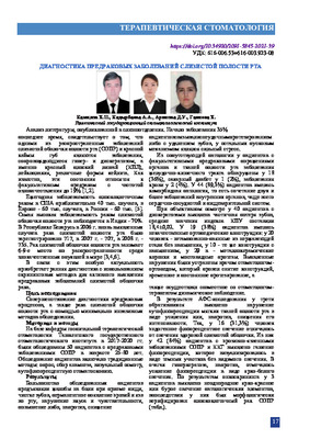 To improve the diagnosis of precancerous processes, as well as cancer of the oral mucosa using minimally invasive examination methods. Material and methods: On the basis of the Department of Hospital Therapeutic Dentistry of the Tashkent State Dental Institute in 2017-2020. 50 patients with precancerous diseases of the oral mucosa at the age of 25-80 years were examined. Results: According to the results of oncological screening in 3 patients, a heterogeneous bright red or brown luminescence of pathological elements was revealed; subsequently, squamous cell carcinoma of the oral mucosa was morphologically verified in them. Conclusions: Early detection of potential precancerous processes allows avoiding interventional diagnostic methods, as well as reducing the risk of developing cancer of the oral mucosa
 -
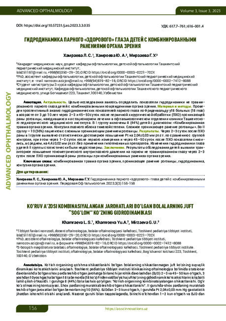
To determine the hydrodynamic parameters of the uninjured fellow eye of children with combined injuries of the organ of vision. A prospective analysis of the hydrodynamic parameters of the fellow eye according to Friedenwald was carried out in 18 patients (18 eyes) aged 3 to 10 years 2–3 and 45–50 days after primary surgical treatment (PSD) of a penetrating wound of the cornea, who were hospitalized in the ophthalmological department of the clinic Tashkent Pediatric Medical Institute. Group I included 8 (44%) children with the following diagnosis: “Combined injury of the organ
of vision. Contusion of the eyeball severe. Complex penetrating wound of the cornea. Group II included 10 (56%) patients with complex penetrating wounds of the cornea. 2–3 days after PST of the wound, group I showed a statistically significant increase in Pt by 2.04±0.03 mm Hg. compared with the control group, while 1–2 days after the first measurement and 45–50 days after PST, the indicators decreased, on average, by 4.4±0.02 mm Hg. without the use of antihypertensive drugs. Changes in the hydrodynamics of the eye in children of group II were not statistically significant. The results of the examination of children revealed a transient increase in tonometric intraocular pressure in the paired uninjured eye 2–3 days after PST of a penetrating wound of the cornea with combined injuries of the organ of vision. -
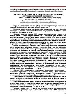 Modern aspects of pathogenesis and features of the course of lesions of the oral mucosa in HIV-associated patientsThis article describes modern aspects of the pathogenesis and characteristics of oral lesion on HIV associated patients. Authors indicated the need for knowledge of the symptoms of oral lesion at different stages of the clinical course of HIV infection. This in turn contributes to the choice of treatment strategy and obtaining long-term positive results m the treatment of oral pathology.
Stomatologiya -
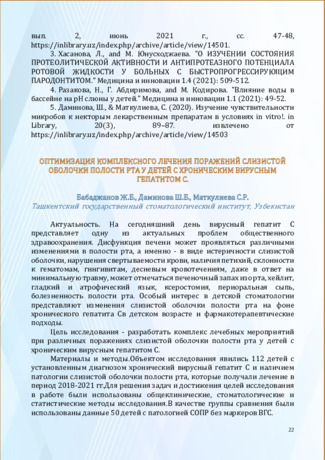 Оптимизация комплексного лечения поражений слизистой оболочки полости рта у детей с хроническим вирусным гепатитом С.
Актуальные вопросы профилактики стоматологических заболеваний и детской стоматологииНа сегодняшний день вирусный гепатит С представляет одну из актуальных проблем общественного здравоохранения. Дисфункция печени может проявляться различными изменениями в полости рта, а именно - в виде истеричности слизистой оболочки, нарушения свертываемости крови, наличия петихий, склонности к гематомам, гингивитам, десневым кровотечениям, даже в ответ на минимальную травму, может отмечаться печеночный запах изо рта, хейлит, гладкий и атрофический язык, ксеростомия, периоральная сыпь, болезненность полости рта. Особый интерес в детской стоматологии представляют изменения слизистой оболочки полости рта на фоне хронического гепатита Св детском возрасте и фармакотерапевтические подходы.
-
 Роль гигиенического ухода полости рта в профилактике кариеса зубов в раннем возрасте.
Actual problems of dentistry and maxillofacial surgery 4Кариес зубов - многофакторное заболевание, в развитии которого важную роль играет высокое качество гигиены полости рта. Знание того, что кариес является динамическим и обратимым процессом, привело к развитию новых технологий, способных выявить кариес на самых ранних стадиях до образования полости. Ежедневная гигиена полости рта с использованием пасты, в состав которых включены фторид и бикарбонат кальция, поможет остановить развитие кариеса зубов и снизить риск появления новых кариозных полостей даже в условиях низкого уровня гигиены полости рта
-
 СОДЕРЖАНИЕ АНТИМИКРОБНЫХ ПЕПТИДОВ В РОТОВОЙ ЖИДКОСТИ У ДЕТЕЙ С ДЕТСКИМ ЦЕРЕБРАЛЬНЫМ ПАРАЛИЧОМУ детей с детским церебральным параличом (ДЦП) показатели заболеваемости кариесом зубов значительно выше в сравнении со здоровым контингентом. Ротовая жидкость оказывает значительное влияние на поддержание гомеостаза полости рта и представляет собой естественную функциональную среду для органов полости рта. Некоторые физико-биохимические показатели ротовой жидкости являются чувствительными индикаторами коморбндной патологии, в частности психоневрологических расстройств, и могуч иметь прогностическое значение для опенки риска развития кариеса. Являясь физиологической «внешней» средой для зубов и других органов полости рта. слюна увлажняет органы полости рта и пищу, осуществляет защитную и трофическую функции. Стабильное и постоянное поступление слюны, которая осуществляет интеграцию мягких и твердых тканей в полости рта. обеспечивает не только поддержание гомеостаза ротовой полости и эффективное удаление эндо- и экзогенных микроорганизмов и их метаболитов, но и постоянное присутствие в различных защитных факторов.[1,2]
Stomatologiya -
 Precancerous lesions of oral mucosa can be considered as diseases, which tend to maligning in various percent.Clinic picture is the same for such diseases. Pathology changes of oral mucosa are absent on early stages, but in malign process there are histological of oral carcinoma examined. Such diseases cause problems affecting social and daily life of the patient. The aim of the study was to improve the diagnosis of precancerous processes, as well as cancer of the oral mucosa, using minimally invasive examination methods. On the basis of the Department of Hospital Therapeutic Dentistry of the Tashkent State Dental Institute, in 2017- 2020, 50 patients with precancerous diseases of the oral mucosa were examined at the age of 25- 80 years. Early detection of potential precancerous processes allows avoiding interventional diagnostic methods, as well as reducing the risk of developing cancer of the oral mucosa.
 -
 COMBINED THERAPY OF CANDIDOSIS OF THE MUCOSA OF THE ORAL CAVITYThe problem of oral candidiasis is relevant both among adults and in children due to frequent recurrence, the presence of a large number of predisposing factors, and the lack of compliance with hygienic measures for oral care. The most common causative agent of oral candidiasis is the fungi C. albicans. The article considers the risk factors for the development of this disease, taking into account age characteristics. Local etiotropic therapy of isolated candida lesions of the oral cavity with the inclusion of broad-spectrum antimycotics is effective in most cases. Preventive measures taken at the end of courses of etiotropic therapy are aimed at reducing the number of relapses of oral candidiasis. The article summarizes the principles of treatment of candidiasis, depending on various factors associated with the course of this disease.
Journal of oral medicine and craniofacial research -
 The oral microflora is one of the most important indicators of the state of the body as a whole. A change in the microflo-ra-organism balance can lead to a change in the local immunity of the oral cavity. According to a number of authors, toothpastes can provide a bacteriostatic and bactericidal effect on gram-positive and gram-negative bacteria and yeast-like funguses. This article introduces the course and results of a stud}', which was based on the determination of bacteriostatic effectiveness of five toothpastes from different production countries.
 -
 Совершенствование качества пародонтологической помощи населению сельской местности
Topical issues of surgical dentistry and dental implantologyПроблема оказания стоматологической помощи сельскому населению считается одним из актуальнейших проблем. Сельскоенаселение имеет меньше возможности получения полномасштабного стоматологического вмешательства. Глобальная программа ВОЗ в области охраны здоровья полости рта проводит свою работу в соответствии со стратегией профилактики хронических болезней и укрепления здоровья. Особое внимание уделяется разработке глобальной политики в области укрепления здоровья полости рта и профилактики болезней полости рта, включая стимулирование разработки и осуществления проектов по укреплению здоровья полости рта и профилактике болезней полости рта на уровне отдельных сообществ при уделении особого внимания социально неблагополучным группам населения (World Health Organization 2009).